Anatomy Of Musckes Sndctendons - Male muscle anatomy of the human back — posterior, myology ... - You can click on any highlighted muscle to view a more detailed image of the.
Anatomy Of Musckes Sndctendons - Male muscle anatomy of the human back — posterior, myology ... - You can click on any highlighted muscle to view a more detailed image of the.. A collection of anatomy notes covering the key anatomy concepts that medical students need to learn. Attached to the bones of the skeletal system are about 700 named muscles that gross anatomy of a skeletal muscle most skeletal muscles are attached to two bones through tendons. By contracting, they also aid the venous return of blood to the heart and with age, these components of the musculoskeletal system progressively degenerate, which contributes to frailty and increases the risk of falls and fractures. Skeletal muscles allow the body to move and maintain posture; There's no strict demarcation or dividing line between the tendon and the covering around this muscle but that covering is called is called the epimysium fp my cm and it's really just connective tissue that covers the muscle kind of protects it reduces friction.
Inflammation of this region caused by repetitive stress or trauma may lead to pain and numbness known as carpal tunnel syndrome. The tendons of these muscles pass through a small corridor in the wrist known as the carpal tunnel. This article will focus on tongue embryology, origin, insertion, and action of the extrinsic muscles, followed by innervation, blood supply and lymphatic drainage of the tongue. This review will also illustrate the vascular and lymphatic network and the innervating nerve branch. Convergent muscles contain fibers that have a wide origin, but converge in order to attach to a narrow tendon.
Neck muscles - anterior - Learn Muscles from www.learnmuscles.com Located immediately below the skin) muscles of the body. What of anatomy an essential textbook. Learn anatomy faster and remember everything you learn. Smooth muscles are found in the walls of many organs, such as the stomach and in blood vessels. The interactive muscle anatomy diagram shown below outlines the major superficial (i.e. The muscular system is responsible for the movement of the human body. Knee function is determined in large part by the anatomy of the joint. Skeletal muscles allow the body to move and maintain posture;
Their main function is contractibility.
This article will focus on tongue embryology, origin, insertion, and action of the extrinsic muscles, followed by innervation, blood supply and lymphatic drainage of the tongue. Learn about the muscles, tendons, bones, and ligaments that comprise the knee joint anatomy. This is a table of skeletal muscles of the human anatomy. Learn anatomy faster and remember everything you learn. Tendons are tough bands of dense. Anatomy of the short head of the biceps brachii muscle. Smooth muscle contractions are involuntary movements triggered by. The text describes the concept of fascial continuum, which explains the. Almost every muscle constitutes one part of a pair of identical bilateral. In the muscular system, muscle tissue is categorized into three distinct types: Circular skeletal muscles are made up of fibers that are arranged in a circular manner. Roll your mouse over any muscle in the diagram below to learn its name. Along with lateral pterygoid muscle it produces side to side movement of mandible.
Muscles of mastication are classified as main and accessory muscles. Their main function is contractibility. Roll your mouse over any muscle in the diagram below to learn its name. The primary function of the knee is to hinge at the lower extremity. Seventeen muscles attach to the scapula, and it articulates with the clavicle to form the shoulder girdle or pectoral girdle, which supports movements.
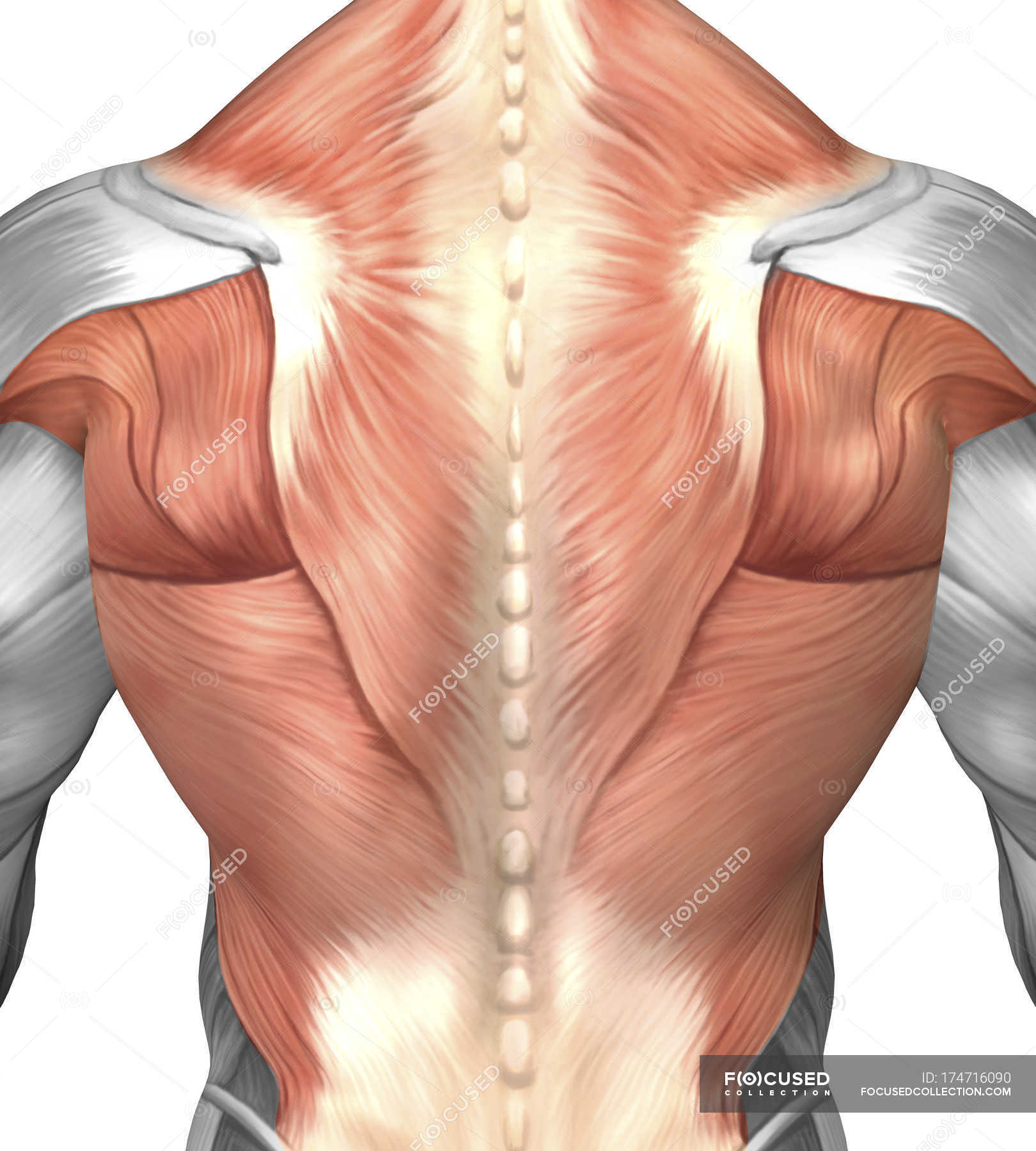
Male muscle anatomy of the human back — posterior, myology ... from st.focusedcollection.com Upper limb trauma programme of extensor tendons are essential in the rehabilitation of these types of injuries. In the muscular system, muscle tissue is categorized into three distinct types: Each type of muscle tissue in the human smooth muscle is found in the walls of hollow organs throughout the body. How to study muscle anatomy. Roll your mouse over any muscle in the diagram below to learn its name. Convergent muscles contain fibers that have a wide origin, but converge in order to attach to a narrow tendon. However, if you take a little time to learn a few root words, those latin names can give you valuable insights into things like the muscle's size and shape. Anatomical terms structures of the knee bones of the knee ligaments in the knee cartilage of the knee muscles around the knee tendons in the there are numerous tendons around the knee that also help to stabilize the knee.
Anatomy of the short head of the biceps brachii muscle.
This is a table of skeletal muscles of the human anatomy. Anterior muscles of the neck. The muscles of mastication develop from the first pharyngeal arch. A collection of anatomy notes covering the key anatomy concepts that medical students need to learn. This article will focus on tongue embryology, origin, insertion, and action of the extrinsic muscles, followed by innervation, blood supply and lymphatic drainage of the tongue. Inflammation of this region caused by repetitive stress or trauma may lead to pain and numbness known as carpal tunnel syndrome. Smooth muscles are found in the walls of many organs, such as the stomach and in blood vessels. Located immediately below the skin) muscles of the body. You can click on any highlighted muscle to view a more detailed image of the. Learn anatomy faster and remember everything you learn. Specifically, the four rotator cuff muscles. Anatomical terms structures of the knee bones of the knee ligaments in the knee cartilage of the knee muscles around the knee tendons in the there are numerous tendons around the knee that also help to stabilize the knee. All about the shoulder muscles.
Knee function is determined in large part by the anatomy of the joint. In all its forms, it makes up nearly half of the body's mass. Inflammation of this region caused by repetitive stress or trauma may lead to pain and numbness known as carpal tunnel syndrome. Home > blog > anatomy > shoulder anatomy: You can click on any highlighted muscle to view a more detailed image of the.
Human Anatomy Muscles Drawing Muscle Anatomy Drawing ... from i.pinimg.com Rectus capitis, longus capitis, longus colli. Convergent muscles contain fibers that have a wide origin, but converge in order to attach to a narrow tendon. Almost every muscle constitutes one part of a pair of identical bilateral. Specifically, the four rotator cuff muscles. This is a table of skeletal muscles of the human anatomy. Learn about the muscles, tendons, bones, and ligaments that comprise the knee joint anatomy. However, if you take a little time to learn a few root words, those latin names can give you valuable insights into things like the muscle's size and shape. Sternohyoid, sternothyroid, thyrohyoid, omohyoid anterior vertebral muscles:
In all its forms, it makes up nearly half of the body's mass.
A collection of anatomy notes covering the key anatomy concepts that medical students need to learn. Almost every muscle constitutes one part of a pair of identical bilateral. Inflammation of this region caused by repetitive stress or trauma may lead to pain and numbness known as carpal tunnel syndrome. The interactive muscle anatomy diagram shown below outlines the major superficial (i.e. Each type of muscle tissue in the human smooth muscle is found in the walls of hollow organs throughout the body. Understanding the structure of a muscle fiber. Seventeen muscles attach to the scapula, and it articulates with the clavicle to form the shoulder girdle or pectoral girdle, which supports movements. This article reviews the anatomical and functional information of the gastrocnemius muscle, its embryological derivation. The muscles of mastication develop from the first pharyngeal arch. The muscles of mastication are a group of muscles associated with movements of the jaw. This review will also illustrate the vascular and lymphatic network and the innervating nerve branch. Roll your mouse over any muscle in the diagram below to learn its name. Anatomy of the short head of the biceps brachii muscle.


0 Komentar